
Electroporation of Xenopus embryo: Gene Delivery into the Primordium Eye at the Neural Plate Stage
Electroporation of Xenopus embryo: Gene Delivery into the Primordium Eye at the Neural Plate Stage
NEPA21 / CUY21 Applications |
|
[Xenopus Embryo] Electroporation of Xenopus embryo: Gene Delivery into the Primordium Eye at the Neural Plate Stage . |
|
|
 |
|
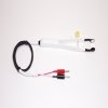
Fig.(1) shows the general setting of the electroporation. Vitelline membrane of the embryo (st.12-13) is usually kept intact. Inject 5-10nl of GFP-mRNA(1µg/µl, 0.05% Fast Green) into intercellular space of upper-few epithelial layers of the neural plate, and immediately after injection (leaving the needle in the embryo) the electric shock (20-22V, 5msecON, 95msecOFF, 10 shocks) is given by the micro-electrode. A: Recognition of the target site by Fast Green just after electroporation. This dye fades away 10-15min later. B: Expression of GFP at the eye 15-20 hours later. C: Detection of GFP protein in the eye vesicle by FITC-conjugated immuno-staining of thin section. The local deliveries of mRNAs such as BMP and Shh result in the perturbation of eye development in addition to the up/down regulation of the eye-marker genes |
|
|
Radioisotope Research Center, Nagoya University; Kazuhito Takeshima *genesis, Volume 33, Issue 2, Pages 81-85, June 2002
|
|
| Contact Us | |
 (Figure 1)