

TiYo LED Autofluorescence Quenching
 |
|
 |
LED Autofluorescence Quenching for CELL and TISSUE STAIN SIGNALS
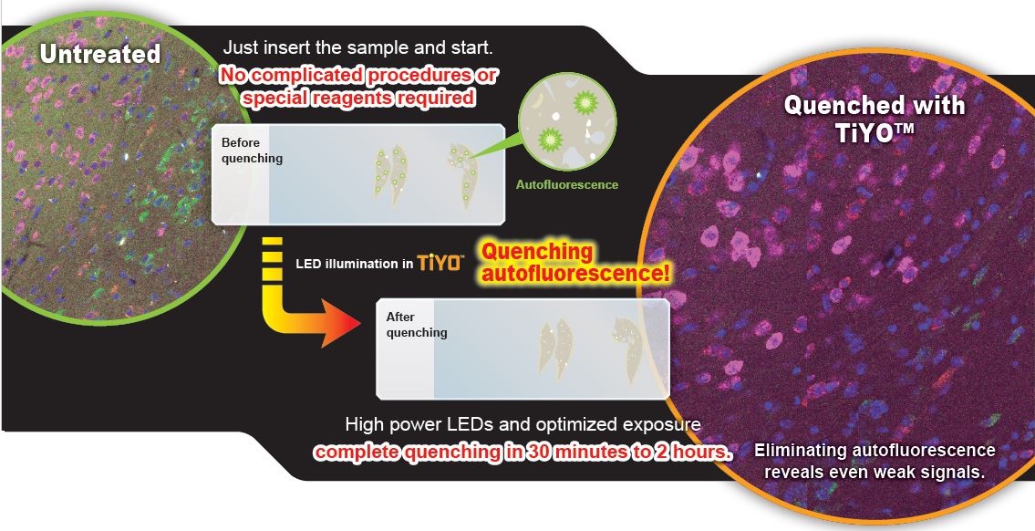
The TiYO™ is a wholly novel, first to market, patented technology that eliminates background autofluorescence signals in cell and tissue stain signals. Noise in tissue observation is caused by substances not related to the staining label but which naturally emits autofluorescence. For example, lipofuscin granules, elastin fibers, vitamin A and other intrinsic molecules that accumulate in cells due to aging emit fluorescence. The autofluorescence generated by such substances present a problem for researchers because it is difficult to distinguish their auto-fluorescence signals from fluorescence signals artificially labeled by staining.
Advantages
- Highly efficient
- Super fast
- One Step Process
- No costly chemical reagents
- DETECT valuable specimens in CLINICAL SETTINGS
- NOT only for imaging technology in histology
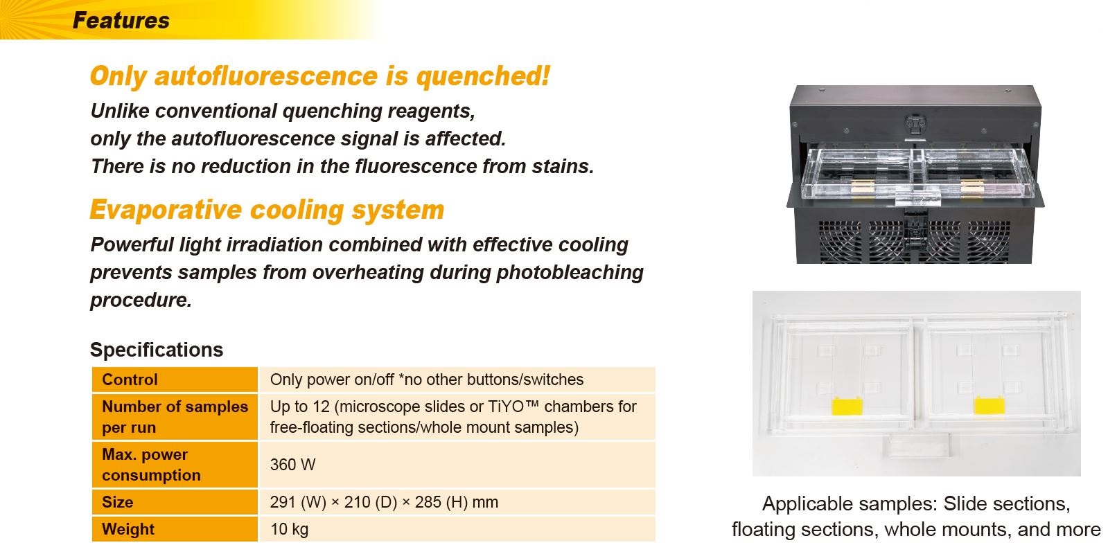
Features
- IDEAL for Highly Autoflourescent species
- ELIMINATE Autofluorescence for microscopy
- Display ONLY Fluorescence Staining
- REVEAL weak stain signals HIDDEN by autofluorescence
- No REDUCTION in stain signal
- No TISSUE DEGENERATION
- No CHEMICAL TREATMENT
- MAXIMISE data from TISSUE SECTIONS
- RE-STAIN a single glass slide MULTIPLE TIMES by TURNING OFF previously stained flourescence
 |
How it works
TiYO™ combines an optimised light irradiation wavelength to photo-bleach unwanted autofluorescence and a novel cooling system that uses the heat of vaporization to remove heat from the surrounding tissue as water evaporates and thus, critically, prevents tissue degeneration due to overheating during the photobleaching procedure.
Compared to competing reagent-based quenching technologies, only the autofluorescence signal is quenched. There is NO REDUCTION in the fluorescence from stains.
IN COMPARISON, competing cooling systems employing Ice and Peltier are TIME CONSUMING and COSTLY and are unable to cool the target tissue in time to prevent tissue degeneration.
One Step Process
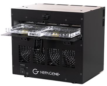
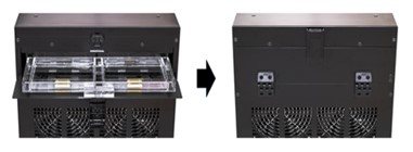
INSERT 1 – 12 samples in the tray and Press START BUTTON
 |
 |
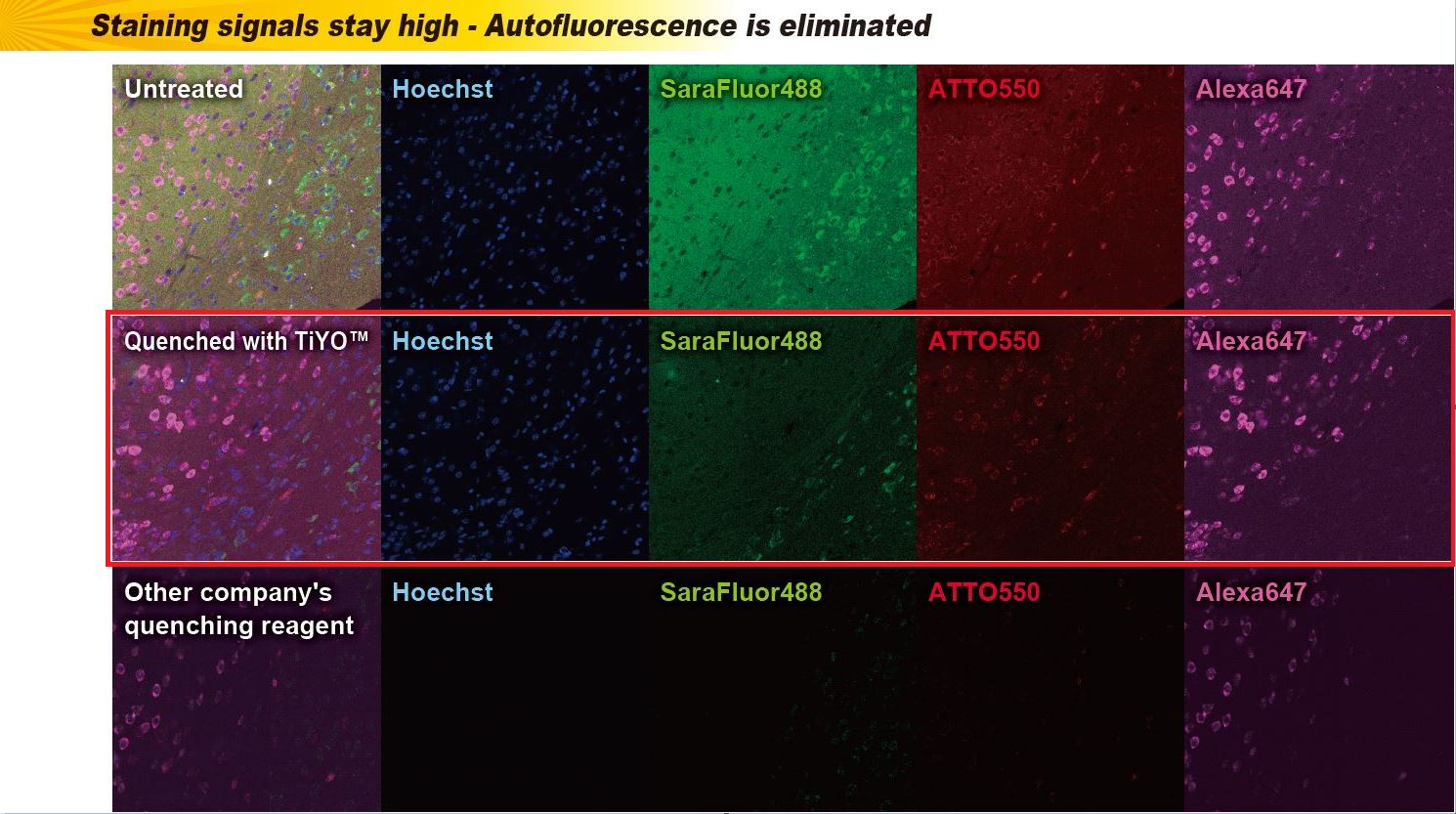
Comparative data for fluorescence staining
Mouse Brain Sections
Without treatment, with TiYO™ pretreatment, and with reagent treatment
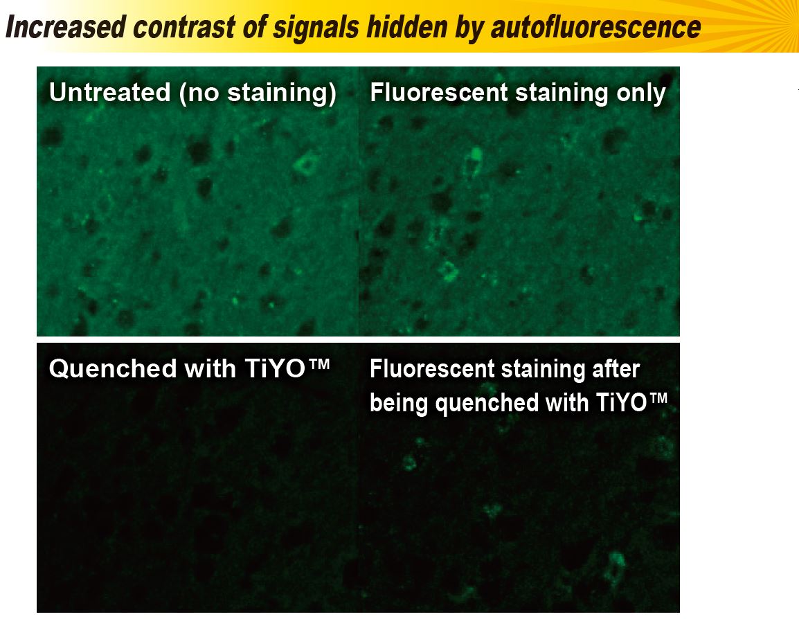
Effective for high autofluorescence tissue types
Chum salmon gill sections are known for their extraordinarily high levels of autofluorescence. A simple two-hour treatment with the TiYO eliminated the autofluorescence.

Autofluorescence of chum salmon gill sections and quenching with TiYO™. The same imaging conditions and contrast settings were used for all images (the outline of the tissue is outlined with translucent white lines).
Courtesy of Dr. Takehiro TSUKADA / Department of Biomolecular Science, Faculty of Science, Toho University
Autofluorescence and quenching treatment with TiYO™ in mouse brain tissue sections. The same imaging conditions and contrast settings were used for all images.
Reference: Fluorescence quenching by high-power LEDs for highly sensitive fluorescence in situ hybridization. Tsuneoka et al. (2022) Front Mol Neurosci
TiYO™ was jointly developed and commercialized using the technology invented by Associate Professor Yosuke Tsuneoka, Faculty of Medicine, Toho University (patent pending: JP-A 2021-008882).
*All features and specifications subject to change without notice.