

Postnatal_Cerebellum_EP_with_the_CUY699P7x6
Postnatal Cerebellum EP using 3 electrodes
There is literature available citing the use of a three-part electrode configuration in conjunction with our CUY21 series electroporator:
High-performance and reliable site-directed in vivo genetic manipulation of mouse and rat brain by in utero electroporation with a triple-electrode probe
Joanna Szczurkowska, Andrzej W. Cwetsch, Marco dal Maschio, Diego Ghezzi, Gian Michele Ratto & Laura Cancedda, Protocol Exchange (2013) doi:10.1038/protex.2013.089, 02 December 2013
Targeted in vivo genetic manipulation of the mouse or rat brain by in utero electroporation with a triple-electrode probe
Joanna Szczurkowska, Andrzej W Cwetsch, Marco dal Maschio, Diego Ghezzi, Gian Michele Ratto & Laura Cancedda, Nature Protocols 11, 399–412 (2016) doi:10.1038/nprot.2016.014, Published online 04 February 2016
For your information, we supply a similar three-part electrode, our CUY699P7x6 electrode.
However, our preferred and recommended electrode configuration for postnatal cerebellum electroporation does not combine three electrodes in an awkward single unit but instead uses our CUY700PL type-electrodes in combination with the following electrodes and cables:
– C115CB cable
– C118 cable
– CUY650P5 or other diameters
– CUY700P4L or other diameters
For your information, Dr. Sakamoto (Columbia University) used our CUY700PL type-electrode configuration (rather than the single CUY699P7x6 unit) to successfully replicated the Cancedda triple-electrode experiment.
Because the results he obtained were from the demonstration we did with him, he said that he needs to further optimize the EP parameters some more:
 |
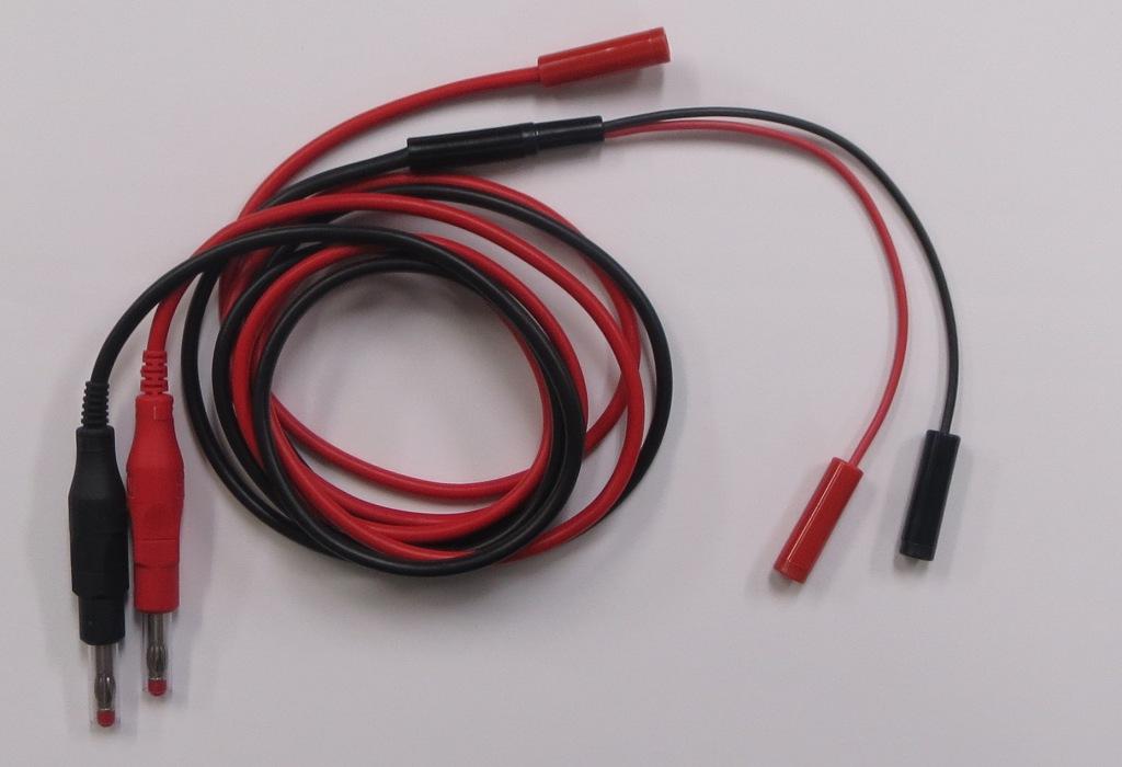
Please note this link to a photo of the C118 cable
One can observer that the black connector-end of the C118 cable fits into the black connector-end of the C115CB Cable.
The remaining red and black ends of the C115CB fit the ends of (say) a CUY650P5/CUY650P10 type-electrode.
The remaining (red) connector of the C118 fit a CUY700P / CUY701P type-electrode.
(The difference between the CUY700 and CUY701 type electrodes is that the latter is square-shape and the former is disk-shaped).
Within the CUY700/CUY701 electrode series, the following options are available:
Possible CUY700P configurations
| Tissue/embryo | CUY700P1L | 1mmφ platinum disk electrode on tip of stick |
| Tissue/embryo | CUY700P2L | 2mmφ platinum disk electrode on tip of stick |
| Tissue/embryo | CUY700P3L | 3mmφ platinum disk electrode on tip of stick |
| Tissue/embryo | CUY700P4L | 4mmφ platinum disk electrode on tip of stick |
| Tissue/embryo | CUY700P5L | 5mmφ platinum disk electrode on tip of stick |
| Tissue/embryo | CUY700P7L | 7mmφ platinum disk electrode on tip of stick |
| Tissue/embryo | CUY700P10L | 10mmφ platinum disk electrode on tip of stick |
| Tissue/embryo | CUY700P20L | 20mmφ platinum disk electrode on tip of stick |
Possible CUY701P configurations
| Tissue/embryo | CUY701P1L | 1mm x 1mm platinum square electrode on tip of stick |
| Tissue/embryo | CUY701P2L | 2mm x 2mm platinum square electrode on tip of stick |
| Tissue/embryo | CUY701P3L | 3mm x 3mm platinum square electrode on tip of stick |
| Tissue/embryo | CUY701P5L | 5mm x 5mm platinum square electrode on tip of stick |
| Tissue/embryo | CUY701P7L | 7mm x 7mm platinum square electrode on tip of stick |
| Tissue/embryo | CUY701P10L | 10mm x 10mm platinum square electrode on tip of stick |
| Tissue/embryo | CUY701P20L | 20mm x 20mm platinum square electrode on tip of stick |
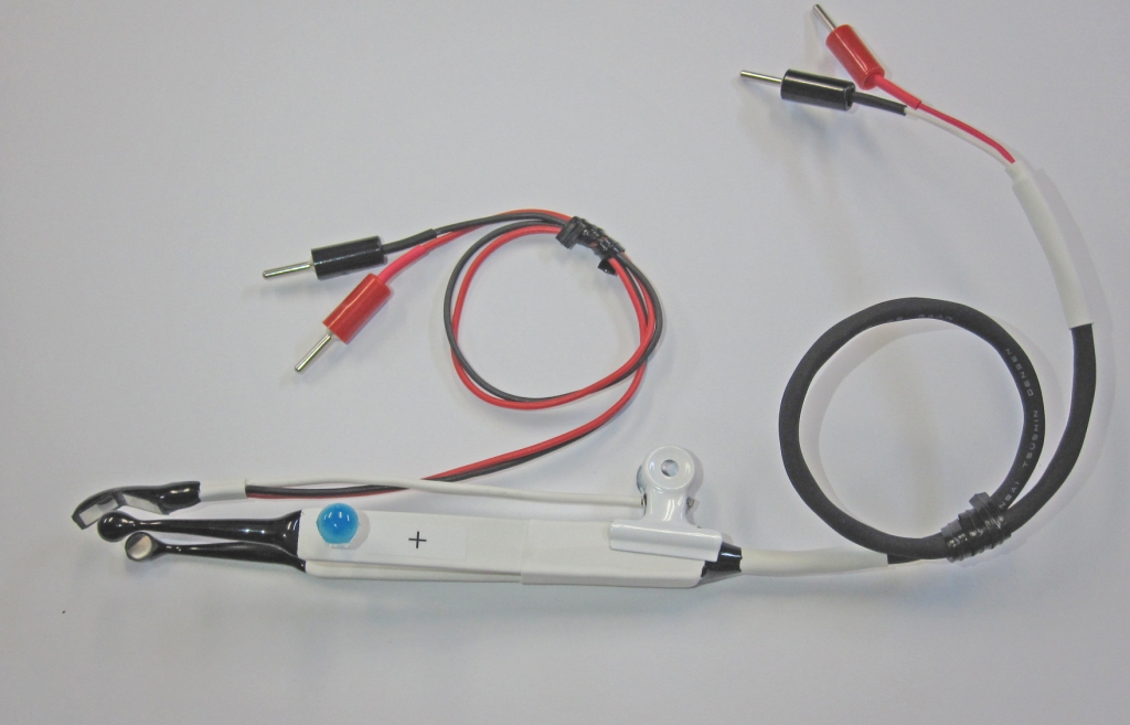
CUY699P7x6 – 3-Part electrode configuration
Three-parts electrodes to electroporate Postnatal Cerebellum
Please note that the CUY669P7X6 configuration is a three-part electrode set-up:
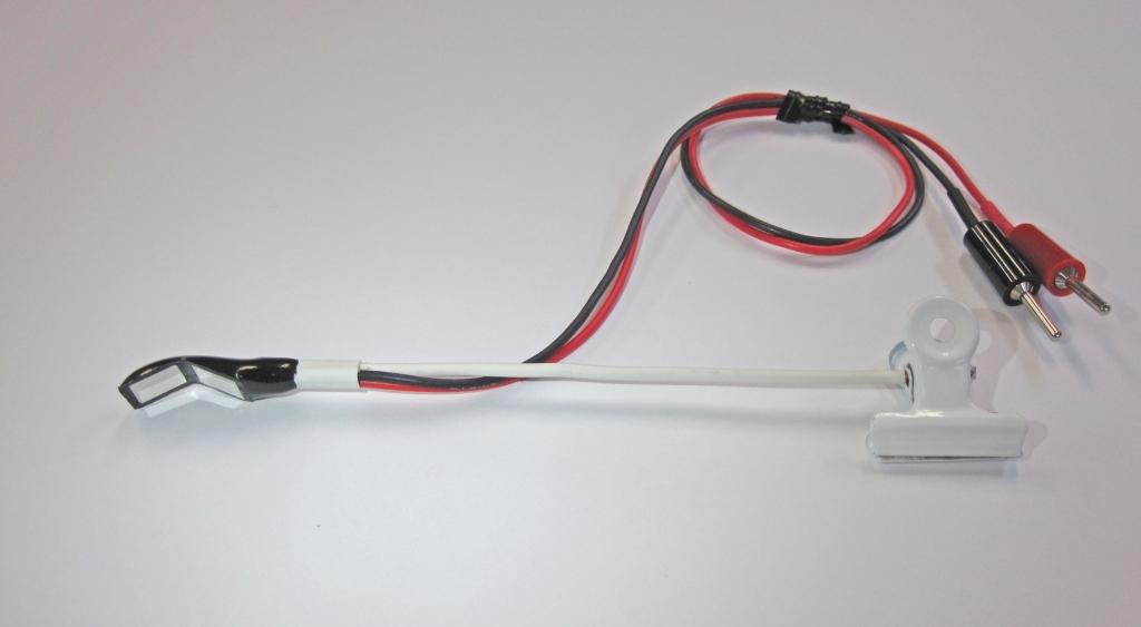
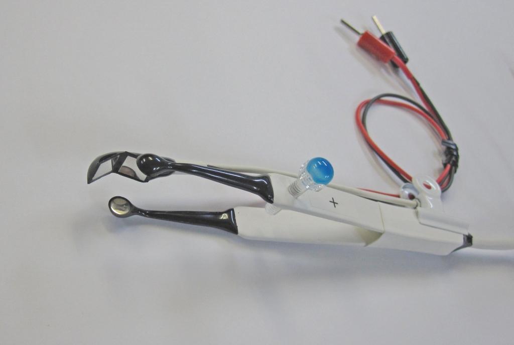
– Photo of the CUY669P7x6
– Photo of the CUY669P7x6 with the CUY650P5 electrode (front angle)
– Photo of the CUY669P7x6 with the CUY650P5 electrode (side angle)
– Diagram for how to set-up and connect the configuration to the NEPA21 electroporator
The CUY669P7X6, it has two electrode plates.
The red electrode cable is connected to the electrode plate with a red-round mark and the black electrode cable is connected to the other electrode plate.
A C118 Connector cable is connected to a tweezers electrode (CUY650P5 or CUY650P10).
Other Relevant Links:
– In Vivo – Cerebellum Pups between 3-8 days old⦁ Ex vivo and In Vivo (Retina)
Postnatal (P0) EP – Mouse Retina Ex Vivo EP
We recommend the CUY650P7 electrode.
For Methodology, please note the publication: Efficient In Vivo Electroporation of the Postnatal Rodent
– Mouse Embryo – Retina/Eye