
Mouse and Rat Tissue and Brain Slice
Mouse and Rat Brain and Tissue Slice Electrode Recommendations:
Brain Slice and Tissue Slice EP
Regarding the correct methodology, please note the article:
Electroporation-mediated Gene Transfer System Applied to Cultured CNS Neurons
Recommended Electrodes
– CUY701P3E and CUY701P2L or
– CUY700P5E and CUY700P2L
NOTE: In the paragraph titled ‘Dissection of hippocampus and electroporation’ please ignore the sentence:
“The dissected hippocampus was placed on the filter with a drop of HBSS and the excess buffer was carefully removed”
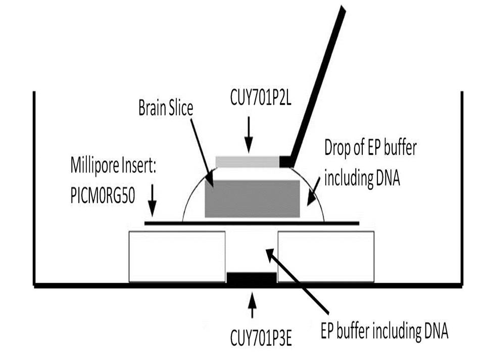
Instead, please follow the figure in this link: Brain Slice and Tissue Slice Configuration Setup
Also, when placing the CUY701P2L electrode on the droplet, please make sure that it does not touch the hippocampus.
To assist in this endeavour, please consider using a micromanipulator.
To view photos of the method used to deliver genes into an entire mouse brain slice with a pair of CUY700P2E and CUY700P2L electrodes, kindly note the following PDF:
– ‘Photos of mouse brain slice electroporation’
If a specific part of the tissue is targeted, it may be appropriate to use a combination of the tungsten-type electrode and the petridish-type electrodes.
The CUY611P3-1 and CUY614T with CUY615 electrodes suffice for such purposes.
Here the tungsten-type needle electrode is used instead of the CUY700P2L electrode recommended above
Because these electrodes are smaller than the CUY702P2L, the transfection area is smaller and more focused.
In Kawabata et al. (2004): Electroporation-mediated gene transfer system applied to cultured CNS neurons
The CUY611P3-1 is used.
We also recommend the CUY614T as it can target a more focused and smaller area.
In Yuzu et al., (2005): The Caudal Migratory Stream: A Novel Migratory Stream of Interneurons Derived from the Caudal Ganglionic Eminence in the Developing Mouse Forebrain
The CUY611P3-1 is used. But the CUY614T is not used.
But this paper is not about Slice EP and the method used is different from our recommended protocol.
The authors also did not use a Millipore Insert.
Brain and Tissue Slice Resource Information
– Application Note 1
– Application Note 2
KnowHow Information
– In Utero Electroporation: Assay system for Migration of Cerebral Cortical Neurons
– In Vivo Electroporation in the embryonic mouse central Nervous System
Re: Postnatal mouse & rat brain EP into the lateral ventricles
Please note the link to a paper on our website (Electroporation and RNA interference in the rodent retina in vivo and in vitro, Matsuda T, Cepko CL.) about postnatal Mouse & Rat EP.
Even though the paper is about the Retina, we believe researchers will find the information useful. Please also note the supporting schematic Electroporation and RNAi in the rodent retina in vivo and in vitro – Fig 8
Please note the following link to an In Utero Resource on our website. Please note that this information is relevant for the ‘Single Step:Single Pulse’ function of the NEAP21
Please also note this link to an In Utero video demonstration of the multiple pulse function of the NEPA21 device
.
NEPA21 – Ex Vivo Publications
– Acute Inhibition of PI3K-PDK1-Akt Pathway Potentiates Insulin Secretion through Upregulation of Newcomer Granule Fusions in Pancreatic β-Cells
– Cortical excitatory neurons become protected from cell division during neurogenesis in an Rb family-dependent manner
– Electroporation-mediated gene transfer system applied to cultured CNS neurons
– Imaging of Insulin Exocytosis from Pancreatic Beta Cells
– In vivo ex vivo retina
– PSD95β regulates plasma membrane Ca(2+) pump localization at the photoreceptor synapse
– The Caudal Migratory Stream, The Caudal Migratory Stream: A Novel Migratory Stream ofInterneurons Derived from the Caudal Ganglionic Eminence in the Developing Mouse Forebrain
See also:
Mouse and Rat Small Tissue Electrode Recommendations and Protocols